
If we can better detect and monitor MS, we can reduce the long-term impact of the disorder.
Research Details
- Multiple Sclerosis Study
- Gisborne & Auckland, NZ
-
Dr Daniel Cornfeld
Dame Helen Danesh-Meyer
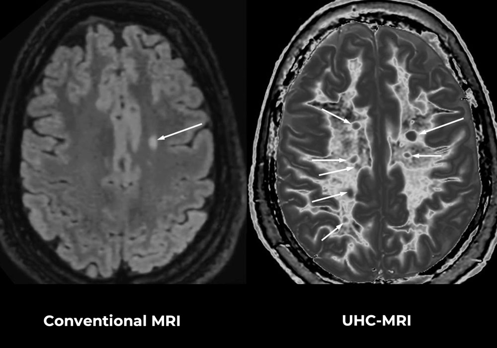
ABOVE: MRI scans of a 32-year-old woman with multiple sclerosis (MS) during a relapse show how advanced imaging can reveal much more disease. A standard scan (left) shows one clear lesion, while the newer UHC-MRI imaging method (right) detects several additional lesions that are not visible on the standard scan. It also shows that much of the surrounding brain tissue is affected, highlighting more widespread disease activity than initially apparent.
Novel MRI Techniques to Enhance MS Lesion Detection and Quantification
Our multiple sclerosis (MS) research is centred around the use of Ultra-High Contrast (UHC-MRI), a novel technique developed by Mātai and collaborators. UHC-MRI enables the detection of subtle lesions and structural changes in the brain, spinal cord, and optic nerves that are not visible using current state-of-the-art imaging.
This advance addresses one of the most critical challenges in MS: the gap between what clinicians can see and what patients experience. Traditional MRI has long focused on visible plaques, yet many symptoms – cognitive, emotional, and physical – cannot be fully explained by these findings alone. UHC MRI reveals changes in both grey and white matter, as well as dynamic tissue alterations during flare-ups. It also highlights previously undetectable processes at lesion boundaries, offering new insight into the active neuroinflammatory mechanisms driving disease progression.
A key innovation underpinning this work is the use of advanced imaging sequences such as directly acquired divided subtracted inversion recovery (dSIR), also known as BLAIR, which exploit tissue properties like T1 relaxation multiple times within a single scan. This allows for sharp contrast at tissue boundaries alongside high spatial resolution – critical for identifying early white matter changes and low-grade inflammation. These capabilities are particularly important for progressive MS, where disease activity often continues without obvious relapses or new lesions on standard imaging.
By enabling earlier and more precise detection of MS lesions, UHC MRI has the potential to shift MS management toward earlier intervention – before irreversible neurological damage occurs. The current study, involving participants both with established MS and those presenting with early symptoms, is specifically aimed at evaluating how sensitive this technology is in detecting disease at its earliest stages.
To participate in this research, see contacts below.
Deep Learning and AI to Accelerate MS Lesion Detection and Quantification
Multiple sclerosis (MS) is a complex immune‑mediated disease with highly variable progression. Identifying patients at risk of worsening disease early is essential for optimising treatment and long‑term outcomes. However, MS often involves subtle, widespread neuroinflammation that is poorly captured by traditional diagnostic tools. This leads to delayed diagnosis, misdiagnosis, and underestimation of disease activity.
Modern MRI has become central to MS evaluation because it can detect lesions, brain tissue damage, and neurodegeneration. However, lesion count alone provides an incomplete picture of disease activity. Lesion shape, spatial distribution, tissue atrophy, and quantitative image features (“radiomics”) add crucial prognostic information.
Using UK biobank data, this research applies artificial intelligence across the full MRI‑to‑prediction pipeline to improve forecasts of disease progression. The models address four clinically important transitions: progression from clinically isolated syndrome to clinically definite MS, from relapsing‑remitting MS to secondary progressive MS, progression independent of relapse activity, and ongoing inflammatory activity seen as new or enlarging lesions. By improving how MRI data are quantified, standardised, and modelled, this work aims to support earlier intervention and more accurate long‑term disease monitoring.
Researchers from Mātai Medical Research Institute in Gisborne are helping develop a new MRI scan method, which they say can detect subtle brain lesions in multiple sclerosis patients not seen on current state-of-the-art scans.
In this accessible presentation for a general audience, Dr Cornfeld explains how the Ultra High Contrast MRI method developed at the Mātai Medical Research Institute could support earlier and more accurate diagnosis of Multiple Sclerosis.