Research Details

  • Ultra-high Contrast MRI (UHC-MRI)
  • Gisborne, NZ
  • Dr Daniel Cornfled
  • research@matai.org.nz

Overview

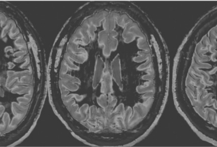
UHC-MRI is a new MRI technique that provides up to 20 times greater visibility of the brain and shows abnormalities which are not currently visible on conventional MRI. The method uses a mathematical framework for increasing the contrast and sensitivity of MRI. This makes it possible to design sequences that will detect very subtle abnormalities.

Once validated UHC-MRI could be applied to images obtained on any clinical scanners across the globe and could be a game changer for neurological conditions like multiple sclerosis, Alzheimer’s disease, stroke, neuroinflammation, brain tumours, and much more.

UHC-MRI was developed by Emeritus Professor Graeme Bydder, Paul Condron, Mātai team members, and other collaborators.
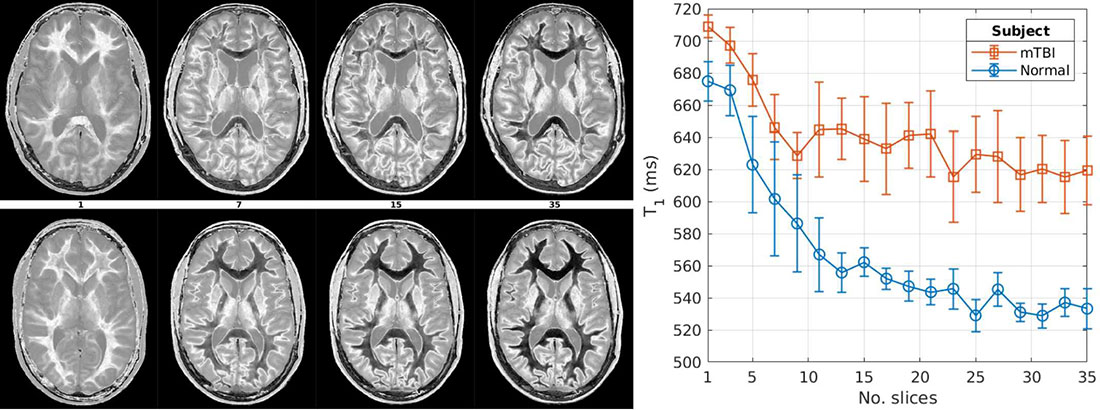
Comparison of UHC images in a patient with chronic mTBI (top) and a healthy volunteer (bottom). The graph shows distinct changes in the white matter relaxation time between the two cases.

Cornfeld, D., Condron, P., Bydder, M., Melzer, T., Pravata, E., Port, J., Holdsworth, S., & Bydder, G. 2025 Dec 31. Ultra-high contrast MRI of the brain and spinal cord using directly acquired and synthetic BipoLAr Inversion Recovery (BLAIR) images. Quantitative Imaging in Medicine and Surgery. [Online] 16:1 

Cornfeld D, Newburn G, Condron P, Emsden T, Bydder M, Scadeng M, Melzer T, Potter L, Holdsworth S, Kwon E, McGeown J, Bydder G. Ultra-high contrast MRI: a new technique for recognizing secondary changes to the brain in patients with persistent symptoms following traumatic brain injury. Neuroradiology. 2026 68(1) 263-276 doi:10.1007/s00234-025-03833-5

McGeown JP, Pedersen M, Mito R, Theadom A, Maller JJ, Condron P, Holdsworth SJ. Neuroimaging correlates of symptom burden and functional recovery following mild traumatic brain injury: A systematic review. NeuroImage: Clinical, 2025; 103910.

Bydder M, Girard OM, Melzer TR, et al. The Role of Incidental Magnetization Transfer in Divided Subtracted Inversion Recovery. NMR Biomed. 2025;38(12):e70165. doi:10.1002/nbm.70165

Condron P, Cornfeld DM, Bydder M, Kwon EE, Whitehead K, Pravatà E, Danesh-Meyer H, Shi C, Emsden TC, Newburn G, Scadeng M, Holdsworth SJ, Bydder GM. Ultra-High Contrast (UHC) MRI of the Brain, Spinal Cord and Optic Nerves in Multiple Sclerosis Using Directly Acquired and Synthetic Bipolar Filter (BLAIR) Images. Diagnostics (Basel). 2025 Jan 30;15(3):329. doi: 10.3390/diagnostics15030329. PMID: 39941259; PMCID: PMC11816702.

Perera MR, Bydder GM, Holdsworth SJ, Handsfield GG. Imaging and image processing techniques for high-resolution visualization of connective tissue with MRI: Application to fascia, aponeurosis, and tendon. Journal of Imaging, 11(2), 43, 2025.

Bydder M, Cornfeld DM, Melzer TR, Condron P, Newburn G, Kwon EE, Tayebi M, Scadeng M, Holdsworth SJ, Bydder GM. Log subtracted inversion recovery. Magnetic Resonance Imaging. 2025;117:110328.

Bydder M, Ali F, Condron P, Cornfeld D, Newburn G, Kwon E, Tayebi M, Scadeng M, Mezler T, Holdworth S, Bydder G. Validation of an ultrahigh contrast divided subtracted inversion recovery technique using a standard T1 phantom. NMR in Biomedicine. 2024;e5269. doi:10.1002/nbm.5269

Condron P, Cornfeld DM, Scadeng M, Melzer TR, Newburn G, Bydder M, Kwon EE, McGeown JP, Handsfield GG, Emsden T, et al. Ultra-High Contrast MRI: The Whiteout Sign Shown with Divided Subtracted Inversion Recovery (dSIR) Sequences in Post-Insult Leukoencephalopathy Syndromes (PILS). Tomography. 2024; 10(7):983-1013. 

Cornfeld D, Condron P, Newburn G, McGeown J, Scadeng M, Bydder M, Griffin M, Handsfield G, Perera MR, Melzer T, Holdsworth S, Kwon E, Bydder G. Ultra-High Contrast MRI: Using Divided Subtracted Inversion Recovery (dSIR) and Divided Echo Subtraction (dES) Sequences to Study the Brain and Musculoskeletal System. Bioengineering (Basel). 2024 Apr 29;11(5):441. doi: 10.3390/bioengineering11050441. PMID: 38790308; PMCID: PMC11118255.

Newburn G, Condron P, Kwon EE, McGeown JP, Melzer TR, Bydder M, Griffin M, Scadeng M, Potter L, Cornfeld D, Holdsworth SJ, Bydder G. Diagnosis of Delayed Post-Hypoxic Leukoencephalopathy (Grinker’s Myelinopathy) with MRI Using Divided Subtracted Inversion Recovery (dSIR) Sequences: Time for Reappraisal of the Syndrome? Diagnostics. 2024;14:418.

Ma YJ, Moazamian D, Port JD, Edjlali M, Pruvo JP, Hacein-Bey L, Hoggard N, Paley MNJ, Menon DK, Bonekamp D, Pravatà E, Garwood M, Danesh-Meyer H, Condron P, Cornfeld DM, Holdsworth SJ, Du J, Bydder GM. Targeted magnetic resonance imaging (tMRI) of small changes in the T1 and spatial properties of normal or near normal appearing white and gray matter in disease of the brain using divided subtracted inversion recovery (dSIR) and divided reverse subtracted inversion recovery (drSIR) sequences. Quant Imaging Med Surg. 2023;13(10):7304-7337.

Newburn G, McGeown J, Kwon E, Tayebi M, Condron P, Emsden T, Holdsworth S, Cornfeld D, Bydder G. Targeted MRI (tMRI) of Small Increases in the T1 of Normal Appearing White Matter in Mild Traumatic Brain Injury (mTBI) Using a Divided Subtracted Inversion Recovery (dSIR) Sequence. OBM Neurobiology. 2023;7(4):201.

Ma Y, Moazamian D, Cornfeld D, Condron P, Holdsworth SJ, Bydder M, Du J, Bydder GM. Improving the Understanding and Performance of Clinical MRI using Tissue Property Filters (TP-filters) and the Central Contrast Theorem, MASDIR (Multiplied, Added, Subtracted and/or Divided Inversion Recovery) Pulse Sequences and Synergistic Contrast MRI (scMRI). Quantitative Imaging in Medicine and Surgery. 2022;12(9):4658-4690.

For more information, contact:

Dr Daniel Cornfeld