
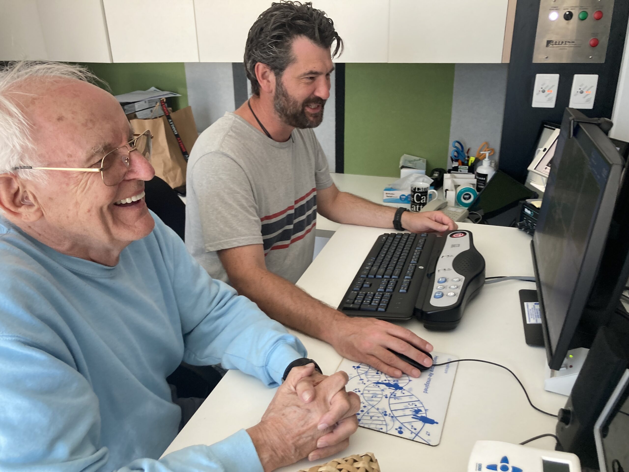
Above: Emeritus Professor Graeme Bydder with Mātai charge technologist Paul Condron seeing UHC MRI in action for the first time. Photo / Mātai Medical Research Institute.
Mātai researchers developing new MRI method
The Gisborne Herald | 10 July 2025
A cutting-edge MRI method developed by the Mātai team and collaborators is capable of detecting subtle brain lesions in MS patients – even in areas that appear normal on standard MRI scans.
When applied in clinical cases, this method revealed abnormal signals during both relapse and remission phases. The ability to detect these changes earlier offers new promise for more precise disease monitoring and could revolutionise how we track MS progression. Ongoing research is underway thanks to support from the New Zealand Multiple Sclerosis Research Trust [tag] and the JN & HB Williams Foundation.
This breakthrough was achieved using a novel Ultra-High Contrast (UHC) MRI technique, which leverages advanced imaging methods that repeatedly exploit tissue properties – particularly T1 relaxation – within a single sequence. One such method, directly acquired divided subtracted inversion recovery (dSIR), is part of a family of BipoLAr Inversion Recovery (BLAIR) sequences, offering both high spatial resolution and enhanced tissue contrast.